
Figure 4a.
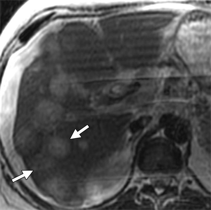
Liver metastases in a 58-year-old man with right colon cancer who had undergone right colectomy 5 years earlier. (a) Axial T2-weighted MR image demonstrates multiple hyperintense hepatic lesions (arrows). Note the perihepatic free fluid, consistent with ascites. (b, c) Axial DW (b = 800 sec/mm2) (b) and ADC (c) images demonstrate diffusion restriction in the periphery of the metastatic deposits as bright signal on the DW image and dark signal on the ADC image (arrows), findings that are consistent with viable tumor. The central portion of the lesion does not show diffusion restriction (ie, relatively higher signal compared with the periphery on the DW image and retention of high signal [T2 shine-through] on the ADC image), findings that are suggestive of necrosis.