
Figure 6.
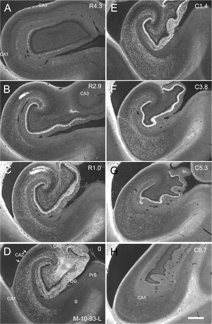
Darkfield photomicrographs of sections of the hippocampal formation arranged from rostral (A) to caudal (H) showing the distribution of BDA labeled fibers and terminals in case M-10-93-L. The injection is located in the proximal portion of CA3 at a mid-rostrocaudal level of the hippocampus (asterisk in D). Note that labeled fibers in CA1, CA2, and CA3 are denser in the superficial portion of stratum radiatum. The CA3 projections to CA1 are more substantial and extensive than the CA3 projections to CA3. Scale bar = 1 mm.