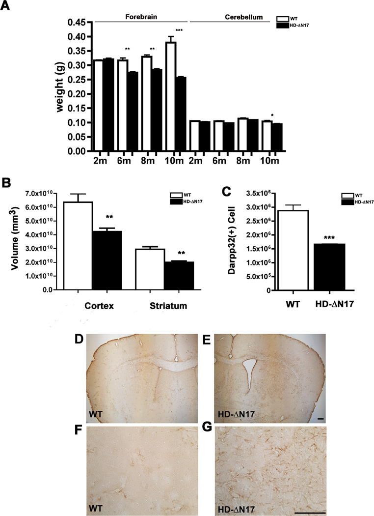
Figure 4. Progressive and Selective Brain Atrophy, Striatal Neuronal Cell Loss, and Gliosis in BACHD-ΔN17 Mice.
(A) Weight of the forebrain and cerebellum of BACHD-ΔN17 and WT littermates was measured at 2, 6, 8 and 10 months of age. Progressive forebrain weight loss is detected in BACHD-ΔN17 mice starting at 6 months of age and progresses between 6m and 10m of age Two-way ANOVA reveals significant differences between HD-AN17 and WT littermates in age and genotype interaction (F=49.94, p<0.0001; genotype: F=173.2, p<0.0001). No weight-loss is detected in the cerebellum until 10m of age. (B) Unbiased stereological measurement of brain regions was performed to measure the cortical and striatal volume in BACHD-ΔN17 mice and WT littermates. (C). Unbiased stereological counting revealed robust loss of Darpp32+ neurons in 10m old BACHD-ΔN17 mice compared to WT controls. (D – G) Reactive astrogliosis is detected in striatum and deep cortical layers of BACHD-ΔN17 mouse brain with GFAP immunohistochemical staining (E, G). No such gliosis is seen in control mouse brain (D, F). Data are shown as mean ± SEM. Student’s t-tests were performed to compare BACHD-ΔN17 and WT samples (*p<0.05; **p<0.01; ***p<0.001). Scale bar = 100 µm. See also Figures S3, S6, S7.

