
Figure 3, 4.
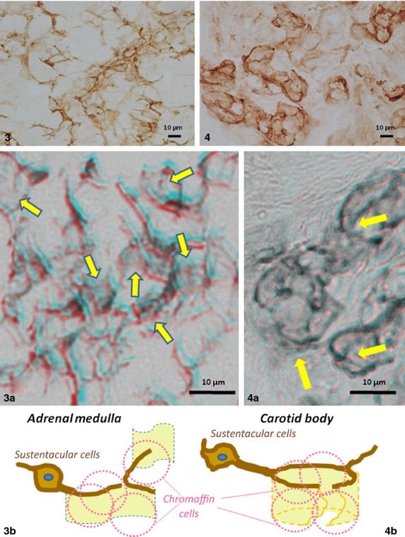
Comparison of light micrographs in morphology of B-FABP-immunostained processes and their lattices in association with weakly immunostained sheets between the adrenal medulla (Fig. 3) and carotid body (Fig. 4) of adult mice. Note open lattices and widely extended sheets of the adrenal sustantacular cells vs. rather close, though incomplete, lattices with sheets in forms of buckets with broken walls of the carotid body sustentacular cells. These differences in architecture of the sheets are shown in 3D light microscopy using red/blue glasses as indicated by arrows in Fig. 3a,b vs. Fig. 4a; and schematically shown in line drawings (Figs 3b and 4b) in which chromaffin cells are shaped in broken pink lines enveloped by sheets.