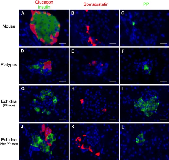
Figure 4.

Immunohistochemical localization of glucagon, insulin, somatostatin and pancreatic polypeptide (PP) in the endocrine pancreas of mouse (A–C), platypus (D–F) and echidna (G–L). (A–C) Consecutive mouse pancreatic sections incubated with the anti-glucagon (A, red), anti-insulin (A, green), anti-somatostatin (B) and anti-PP (C) antibodies. (D–F) Consecutive platypus pancreatic sections incubated with the anti-glucagon (D, red), anti-insulin (D, green), anti-somatostatin (E) and anti-PP (F) antibodies. (G–L) Consecutive echidna pancreatic sections incubated with the anti-glucagon (G, J, red), anti-insulin (G, J, green), anti-somatostatin (H, K) and anti-PP (I, L) antibodies. In all three species, glucagon, insulin, somatostatin and PP immunoreactivities were present on different cell populations. Scale bar: 20 μm.
