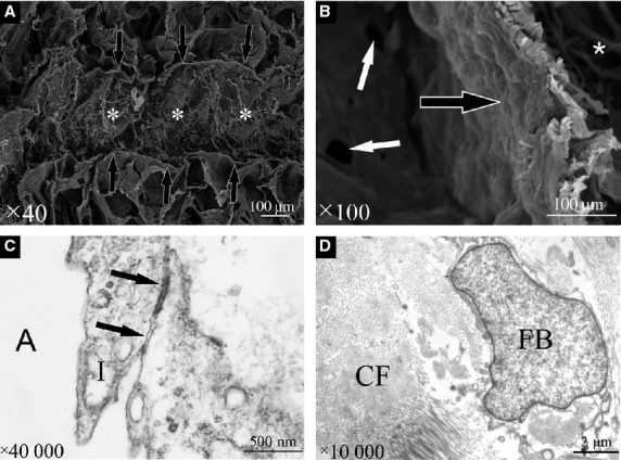
Figure 3.

Scanning electron microscopy photographs of the intersegmental septum. (A) The intersegmental septum consists of three layers, including the superficial layers (black arrows) and deep layer (white asterisk). (B) The superficial layer (black arrow) is integral, and no Kohn's pores were identified. Kohn's pores (white arrows) are only seen on the alveolar wall between two alveoli. The deep layer (white asterisk) contains collagen fibres. Transmission electron microscopy photographs of the intersegmental septum are shown. (C) Type I cells constitute the alveolar wall and contain tight junctions (black arrows). (D) The deep layer is composed of collagen fibrils and fibroblasts. A, alveoli; CF, collagen fibres; FB, fibroblasts; I, type I cell.
