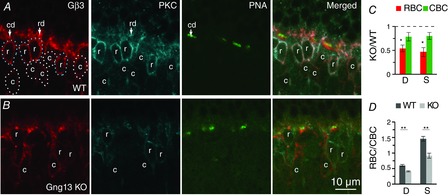
Figure 8.

Absence of Gγ13 reduces Gβ3 staining in RBCs more than in ON CBCs
Triple labelling for Gβ3, PKC and PNA in WT (A) and Gng13 KO (B) retinas. PNA staining locates cone bipolar dendritic tips (cd); the rest of the OPL staining was considered as rod bipolar dendrites (rd). Somas stained for PKC are rod bipolar somas (r); somas stained for Gβ3 and not for PKC are cone bipolar somas (c). ROIs for rod bipolar somas (dotted outlines) and ON cone bipolar somas (white dotted outlines) are shown (A). Note that, in WT mice, the rod bipolar somas display brighter Gβ3 staining than the cone bipolar somas and, in KO mice, the cone bipolar somas have brighter staining. C and D, quantitative analysis of staining intensity. Average intensities per pixel were measured for the four different ROIs (rod bipolar dendrites, cone bipolar dendrites, rod bipolar somas and cone bipolar somas). C, staining intensities in dendrites (D) and somas (S) of KO RBCs (but not ON CBCs) are significantly different from that in WT cells. D, same data set as in C recalculated to show the average ratio (RBC/CBC) of Gβ3 staining intensity in the cell dendrites (D) and somas (S) in WT and KO retinas. For both dendrites and somas, the differences between these ratios for WT and KO are highly significant (4 sets of animals).
