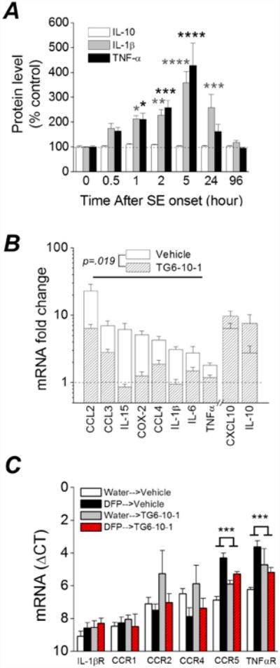
Figure 5. Induction of inflammatory cytokines and receptors 4 days after DFP induced status epilepticus in rats is attenuated by TG6-10-1.

A, changes in IL-1β, TNFα and IL-10 protein in the forebrain at various times after DFP exposure measured by ELISA. (* p < .05, ** p < .01, *** p < .001, **** p < .0001, two-way ANOVA with Dunnett's post hoc test compared to time 0; n = 3-5 rats at each time point). B, change in abundance of 10 inflammatory mediator mRNAs from the forebrain of rats 4 d after injection with water or DFP to induce status epilepticus. Post-DFP treatment was 6 doses of TG6-10-1 (n = 8 rats) or vehicle (n = 9 rats). Following DFP induced status epilepticus, the mRNA fold change for 8 mediators as a group was significantly reduced by TG6-10-1 compared to vehicle (p = .019, paired t test). C, qRT-PCR was also performed to assess the abundance of 6 inflammatory cytokine and interleukin receptors (Table 2) (n = 8 rats per treatment group). The mRNA fold change for 2 of the receptors (TNFαR and CCR5) was significantly reduced by TG6-10-1 compared to vehicle (p < .001, one-way ANOVA with posthoc Bonferroni).
