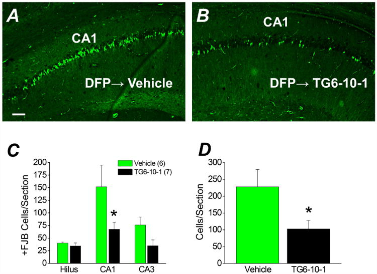
Figure 7. Neuroprotection by TG6-10-1 following DFP-induced status epilepticus.

Representative images of FluoroJade B staining in hippocampal sections (8 μm) in the CA1 region for rats treated with 6 doses of vehicle (n = 6 rats) (A) four days after DFP-induced status epilepticus and rats injected with 6 doses of TG6-10-1 (n = 7 rats) (B). Images were taken at a total magnification of 100×. The images are representative of 8 sections per rat. Scale bar, 100 μm. C, the average number of injured neurons per section in three hippocampal regions of rats treated with 6 doses of vehicle (n = 6 rats) and rats injected with 6 doses of TG6-10-1 (n = 7 rats) four days after DFP-induced status epilepticus. (* p < .05 in CA1, one-way ANOVA with posthoc Bonferroni). D, the average number of injured pyramidal neurons from CA1 and CA3 combined per section. (* p < .05, t test). +FJB, positive FluoroJade B.
