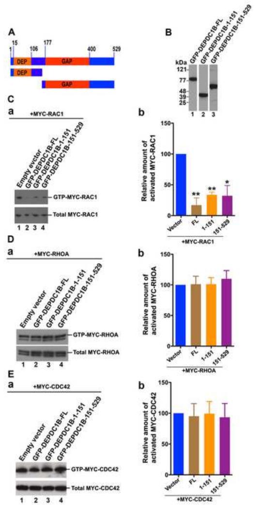
Figure 5. Exogenous expression of DEPDC1B suppresses RAC1 activation.
(A) Schematic of DEPDC1B polypeptides that were used in (B), (C), (D), and (E). The number above the diagram indicates the amino acid residues. (B) Immunoblots showing that GFP-tagged DEPDC1B full-length (lane 1) and fragments (lanes 2 and 3) are expressed in transfected HeLa cells. (C-E) Plasmids encoding MYC-RAC1 (C), MYC-RHOA (D), or MYC-CDC42 (E) were co-transfected into HeLa cells with empty vector or plasmids encoding GFP-DEPDC1B-FL, GFP-DEPDC1B-1-151, or GFP-DEPDC1B-151-529. The transfected cells were subjected to PBD pull-down assays to assess the levels of activated MYC-RAC1 (C) and MYC-CDC42 (E) or subjected to RBD pull-down assays to assess the levels of activated MYC-RHOA (D). Typical immunoblots for the activation assays of RAC1 (Ca), RHOA (Da), and CDC42 (Ea) were shown. Relative amount of activated MYC-RAC1 (Cb), MYC-RHOA (Db), and MYC-CDC42 (Eb) in each group of transfected cells were quantitated based on three independent experiments. The error bars indicate standard deviation around the mean. In (Cb), (Db), and (Eb), FL, 1-151, and 151-529 represent GFP-DEPDC1B-FL, GFP-DEPDC1B-1-151, and GFP-DEPDC1B-151-529, respectively. **, p<0.01; *, p<0.05.

