Figure 3.
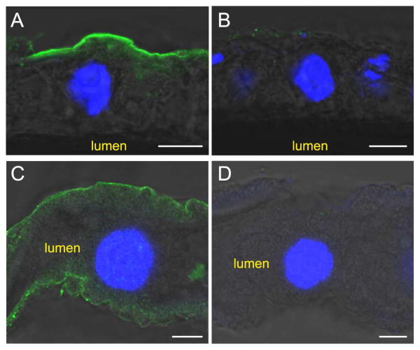
Immunolocalization of AgMCO1 in adult female mosquitoes. Cryosections of midguts (A and B) and unsectioned Malpighian tubules (C and D) were immunostained with AgMCO1 antiserum (A and C) or pre-immune serum (B and D). Anti-MCO1 antibodies were detected with Alexa Fluor 488-conjugated anti-rabbit IgG antibodies (green). Nuclei were stained with DAPI (blue). AgMCO1 immunoreactivity was detected along the basal surface of the epithelial cells of the midgut and Malpighian tubules (A and C). No signal was observed in samples incubated with preimmune serum (B and D). (Scale bars: 10 μm)

