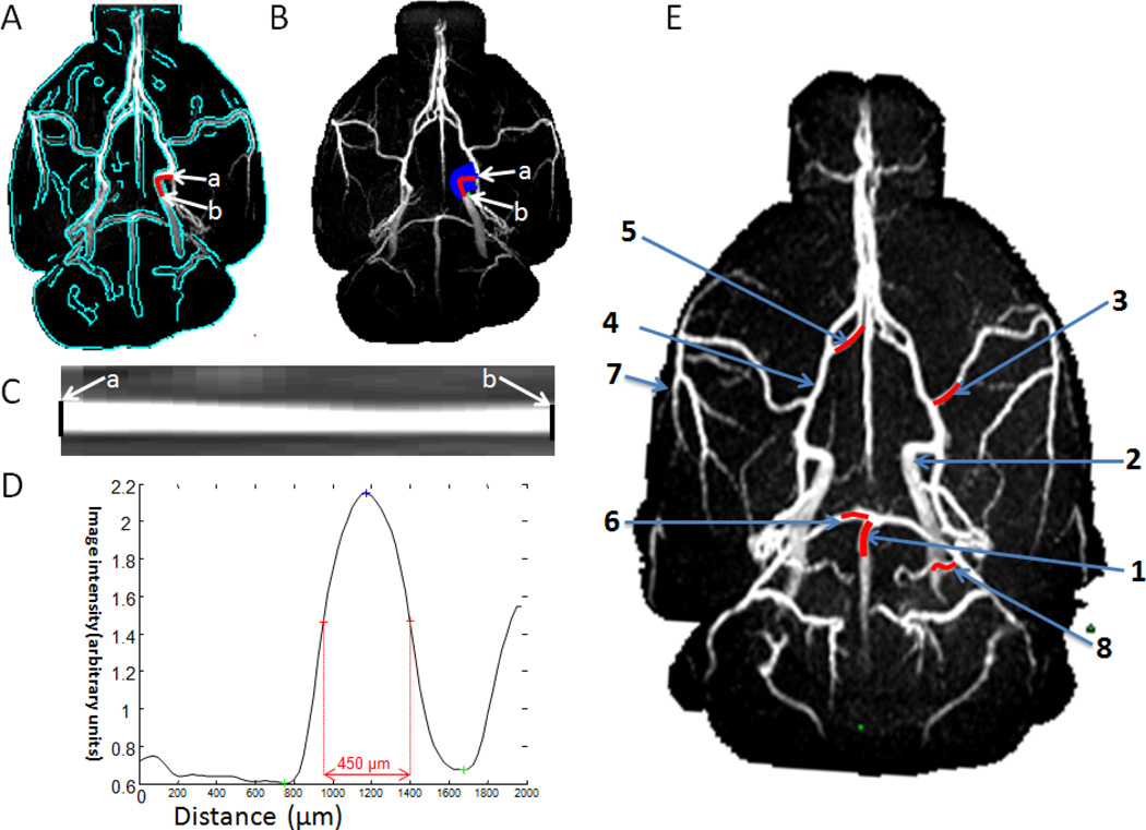
Figure 3.
Automated determination of MRA vessel lumen diameter. (A) The vessel was segmented by using edge-detection technique (green trace). (B) Radial projections perpendicular to the vessel boundary were obtained. The lumen diameter of the ICA (an example) was quantified from point a to point b. The vessel was displayed in (C) image and (D) intensity profile format were flattened. The lumen diameter was defined as the full-width at half-height. (E) The 2 mm segments (red lines) along the artery indicated the lengths over which the diameter was measured. 1: basilar artery (BA), 2: internal carotid artery (ICA), 3: middle cerebral artery (MCA), 4: anterior cerebral artery (ACA), 5: azygos artery, 6: posterior cerebral artery (PCA), 7: pial branch of MCA; 8: pial branch of PCA.

