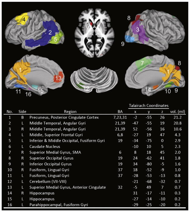
Fig. 3.
Functional regions of interest (fROIs) generated from a disjunction mask derived from the Low Risk and APOE ε4 groups at baseline (0 months), 18 months, and 57 months (see Methods). fROI region numbers correspond to numbers in Tables 3–5. BA= Brodmann’s areas; R= right, L= left, B = bilateral; SMA = supplementary motor area.

