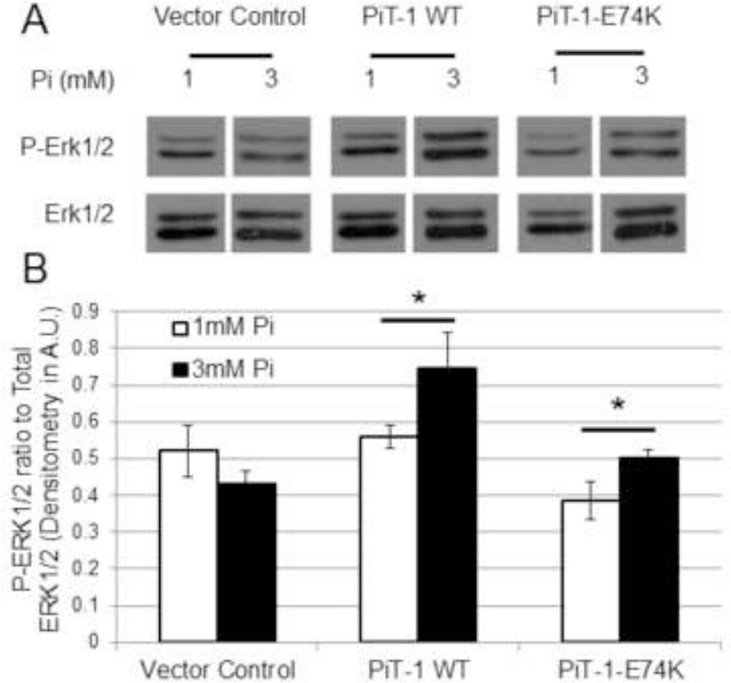
Figure 5. Elevated Pi induced ERK1/2 phosphorylation through Pi transport-independent PiT-1 function.
(A) ERK1/2 phosphorylation was induced in PiT-1 ΔSM VSMCs expressing vector control, PiT-1 WT, or PiT-1-E74K with incubation in 1.0 mM or 3.0 mM Pi for 15 minutes. P-ERK1/2 and total ERK1/2 were visualized by western blot analysis. (B) Densitometry quantification of three independent experiments shows the ratio of P-ERK1/2 to Total ERK1/2. Data presented as a representative image (A) or mean ± S.D., n = 3 for all data points (B). Statistically significant differences between two independent means are indicated by * = P<0.05 as measured by student t-test.

