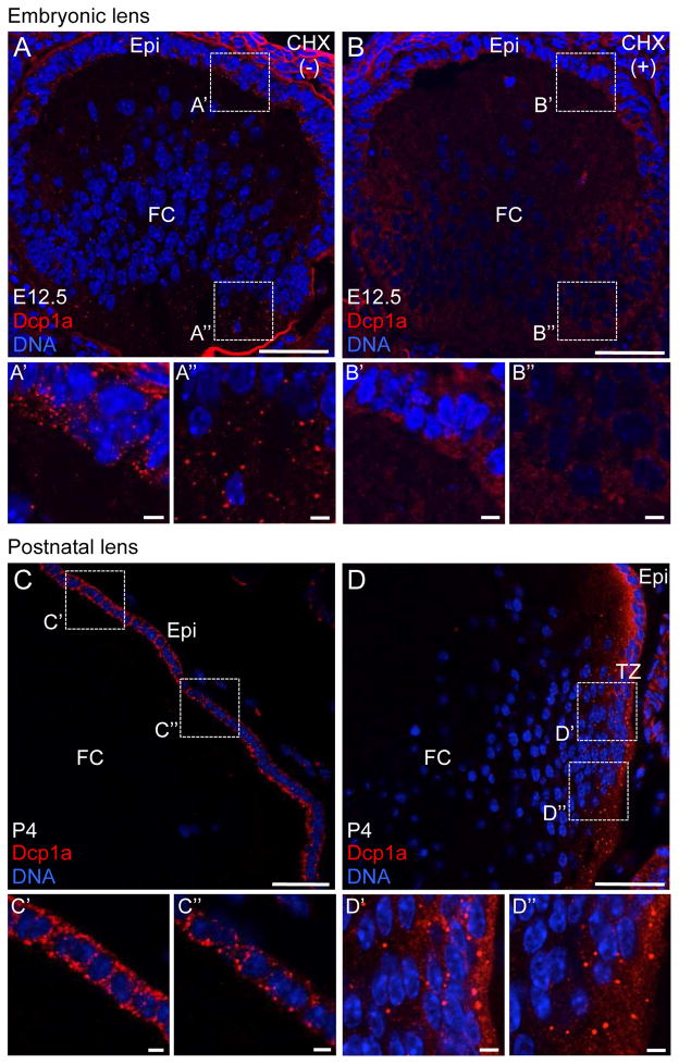
Figure 5. Embryonic and early postnatal mouse lens harbors cytoplasmic Processing bodies.
(A) Immunostaining by Dcp1a, a Processing body (P-body) marker, demonstrates that mouse lens at stage embryonic day 12.5 (E12.5) harbors P-bodies in both lens anterior epithelium (Epi) (A′) as well as in fiber cells (FC) (A″). (B) In presence of translational elongation inhibitor cycloheximide (CHX), these cytoplasmic structures can be disassembled in both Epi (B′) and FC (B″), indicating them are bona fide P-bodies. (C) Mouse lens at postnatal day 4 (P4) exhibits P-bodies in the Epi (C′) and (C″) but not in FC. (D) P4 mouse lens also exhibits P-bodies in the transition zone (TZ) (D′) and (D″). Similar results were obtained using Ddx6 as a P-body marker (data not shown). Dotted line box indicates area selected for representation at high magnification. Scale bar in A–D is 50 μm and in A′, A″, B′, B″, C′, C″, D′, D″ is 5 μm.

