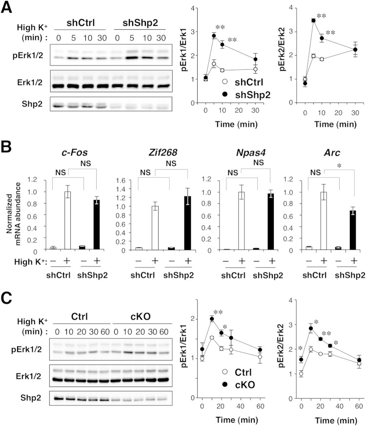
FIG 5.
Effects of Shp2 ablation on membrane depolarization-induced Erk activation and IEG expression. (A) Immunoblot analysis of Erk1/2 phosphorylation in cultured mouse cortical neurons infected with a lentivirus encoding Shp2 or control shRNAs and incubated in culture medium containing 25 mM KCl for 0, 5, 10, or 30 min. Quantitative data are expressed relative to the value for shCtrl-treated cells at time zero. (B) Quantitative PCR analysis of IEG expression in cultured neurons infected as described for panel A and exposed to normal culture medium (5 mM KCl) or medium containing 25 mM KCl for 60 min. Data are expressed relative to the value for shCtrl-treated cells exposed to a high K+ concentration. (C) Immunoblot analysis of Erk1/2 phosphorylation in synaptosomes prepared from the brains of Shp2flox/flox (control [Ctrl]) or Shp2 cKO mice at 43 to 46 weeks of age and exposed to medium containing 25 mM KCl for the indicated times. Quantitative data are expressed relative to the control value at time zero. All quantitative data are the mean value ± SEM of three independent experiments (A, C) or six culture dishes in three independent experiments (B). *, P < 0.05; **, P < 0.01 (Student's t test) for comparisons between shCtrl and shShp2 neurons (A, B) or between Ctrl and Shp2 cKO synaptosomes (C); NS, not significant.

