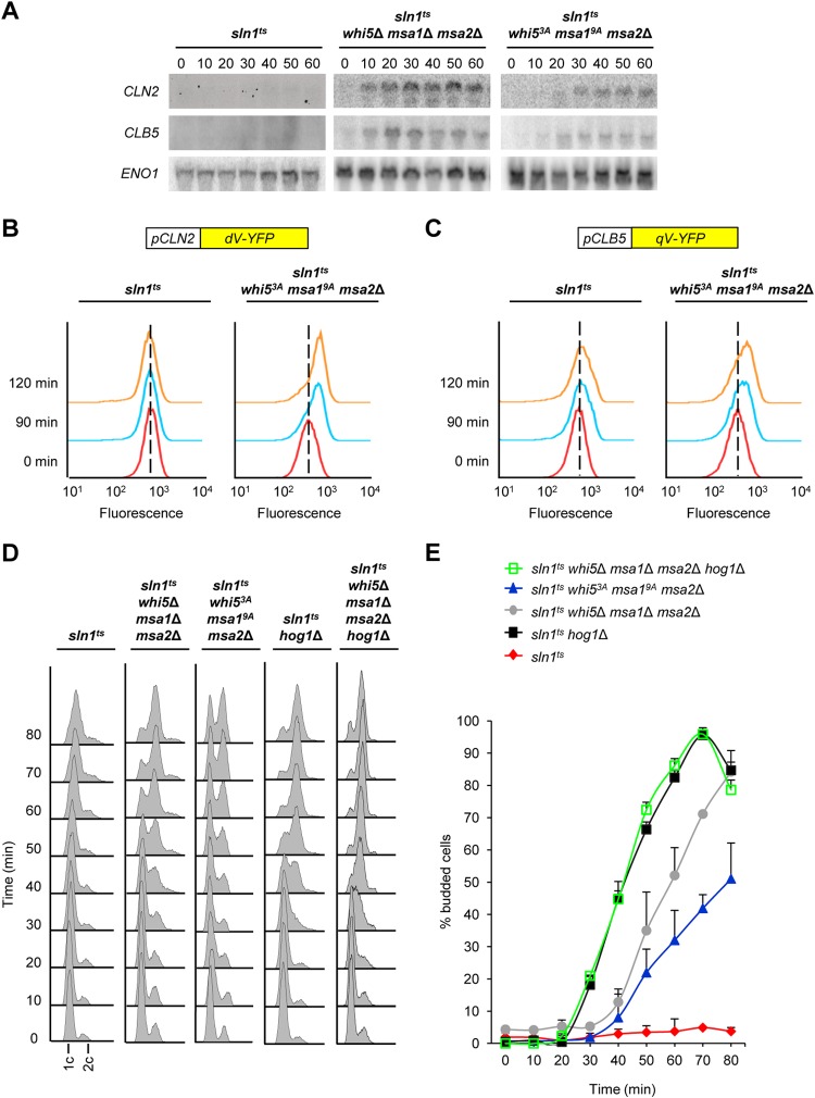
FIG 7.
Phosphorylation of Whi5 and Msa1 by Hog1 prevents G1 arrest. (A) The whi5 msa1 msa2 triple mutant suppress the delay of cyclin expression upon Hog1 activation. The strains indicated were synchronized with α-factor and released at 37°C into YPD medium. RNA was extracted from samples taken at the times indicated after release and analyzed by Northern blotting with radiolabeled probes specific for the CLN2, CLB5, and ENO1 mRNAs. (B, C) Downregulation of both the CLN2 and CLB5 promoters depends on Hog1 phosphorylation of Whi5 and Msa1. sln1ts and sln1ts whi53A msa19A msa2Δ mutant cells were transformed with a fluorescent reporter system for analysis of CLN2 (B) or CLB5 (C) promoter activity. Fluorescence-positive cells were synchronized with α-factor and released at 37°C into YPD medium, and promoter-associated fluorescence was analyzed by flow cytometry in G1-synchronized cells (red lines) or at 90 min (blue lines) or 120 min (orange lines) after release. Each line in the histogram represents the fluorescence distribution from 20,000 cells. (D) G1 arrest depends on the phosphorylation of Whi5 and Msa1 by Hog1. The strains indicated were synchronized with α-factor and released into YPD medium at 37°C. Cell samples were fixed every 10 min after release, and their DNA content was measured by flow cytometry. (E) The Hog1-dependent budding block is mediated by Whi5, Msa1, and Msa2. The same cells as in panel D were microscopically analyzed, and budded cells were counted at the times indicated after release. sln1ts, sln1ts hog1Δ, sln1ts whi5Δ msa1Δ msa2Δ, sln1ts whi53A msa19A msa2Δ, and sln1ts whi53A msa19A msa2Δ hog1Δ mutant cells were analyzed. At least 100 cells of each strain were counted at each time point. Bars represent the averages ± the standard deviations from three independent experiments.

