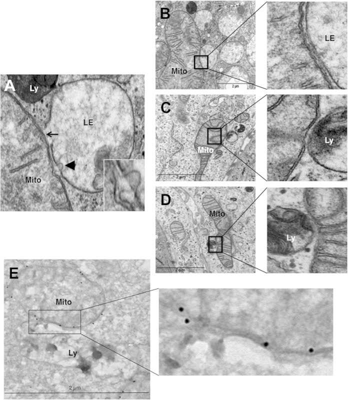
FIG 4.
Hypoxic LS147 cells show close interactions between mitochondria and late endosomes and lysosomes. (A) A representative image from an electron micrograph of an LS174 cell showing an interaction between a mitochondrion (Mito) and a late endosome (LE) showing membrane rupture (arrow) and a late endosome with a lysosome (Ly) showing membrane rupture (arrowhead; see the inset). (Inset) Enlarged view of a late endosome showing blebbing at the mitochondrion-late endosome contact site. (B to D) (Left) Representative electron micrographs of LS174 cells showing the interaction of a mitochondrion with late endosomes and lysosomes. (Right) Enlarged views of the interaction sites. (E) (Left) Representative electron micrograph of LS174 cells showing immunogold-stained VDAC, allowing detection of mitochondria making contact with a lysosome. (Right) Enlarged view of the contact site.

