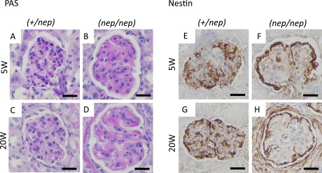
Fig. 1. .

Histological images of glomeruli and podocyte-specific protein at 5 and 20 weeks of age. (A–D) PAS stain, (E–H) nestin stain. (A, E) 5W of heterozygotes, (B, F) 5W of homozygotes, (C, G) 20W of heterozygotes, (D, H) 20W of homozygotes. Bars=20 μm.
