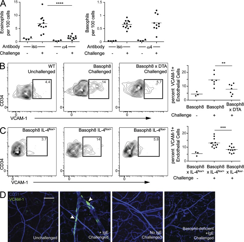
Figure 4.
Basophil-derived IL-4 modulates endothelial cell expression of VCAM-1. (A) Basoph8 mice were sensitized with anti-DNP IgE, and 1 d later, the mice received anti-α4 antibody by intraperitoneal injection. Immediately after antibody administration, DNFB was applied to the ear skin of sensitized mice. Accumulation of eosinophils (left) and basophils (right) in ear skin was assessed 3 d after challenge. Results were pooled from three independent experiments with at least six mice in each challenged group. (B) Basoph8 mice were sensitized with anti-DNP IgE, and 1 d later, the mice were challenged by topical application of DNFB. 2 d after challenge, surface VCAM-1 expression on ear endothelial cells (CD45−CD34+ESAM-1+) from basophil-sufficient (middle) and -deficient (right) mice was examined. The left panel depicts baseline VCAM-1 staining in unchallenged mice. Gates were set against an isotype control–stained sample. The graph on the right represents results pooled from three independent experiments with at least six mice in each challenged group. Littermate basophil-sufficient and -deficient animals were used in this experiment. (C) Basoph8 × IL-4flox/+ (middle) and Basoph8 × IL-4flox/− (right) littermates were sensitized, challenged, and analyzed as in B. The contour plots are representative of the results, with the graph on the right depicting results pooled from two independent experiments with at least six mice in each challenged group. (D) WT and basophil-deficient (Basoph8 × DTA) mice were sensitized and challenged as in B. 2 d later, mice received an i.v. injection of PE-conjugated anti–VCAM-1 and APC-conjugated anti-CD31 antibody. 10 min after antibody injection, whole mount micrographs were obtained using laser-scanning confocal microscopy. Each image stack was then transformed into a maximum intensity projection with VCAM-1– and CD31-stained endothelium. The images were arranged as follows (from left to right): unchallenged WT mice, IgE-sensitized/challenged WT mice, nonsensitized/challenged WT mice, or sensitized/challenged basophil-deficient mice. Arrowheads denote VCAM-1–positive patches. The WT mice were bred independently of the basophil-deficient mice. The image is representative of two separate experiments with at least two mice in each group. Bar, 200 µm. (A–C) Horizontal bars denote mean. P-values were determined by ANOVA: **, P < 0.01; ***, P < 0.001; ****, P < 0.0001.

