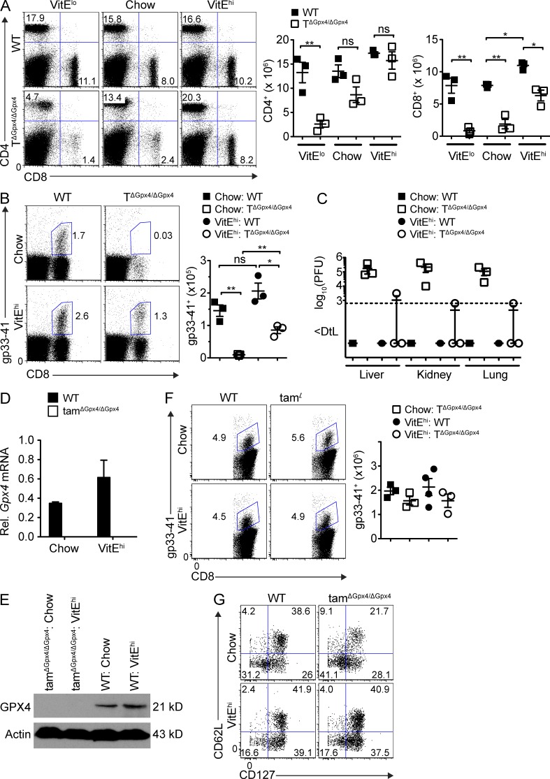
Figure 6.
Vitamin E rescues CD8+ T cell defect and restores viral clearance. Mice fed with vitamin E low (VitElow; <10 mg/kg), chow (50 mg/kg), or vitamin E high (VitEhi; 500 mg/kg) for 3 wk. (A) Flow cytometry (left) and absolute number of splenic CD4+ (middle) or CD8+ T cells (right) in WT and TΔGpx4/ΔGpx4 mice after 3 wk of diet supplementation (n = 3 per group of 6-wk-old mice). (B) Flow cytometry (left) and total number (right) of gp33-41 tetramer specific CD8+ T cells in the spleen (n = 3 per group) at 7 dpi with LCMV WE 200 pfu. (C) Viral titers of LCMV (WE 200 pfu) infected mice in the liver, lung, and kidney (n = 3 per group) at 7 dpi. (D and E) Quantification of Gpx4 mRNA levels in splenic MACS sorted CD8+ T cells. Expression was normalized to G6pdx mRNA levels. (E) Western blot of GPX4 expression withactin as loading control in splenic MACS sorted CD8+ T cells. (F) Flow cytometry of splenocytes infected with LCMV WE (200 pfu), followed by Gpx4 deletion with tamoxifen (2 mg for 2 d) and infection with L. monocytogenes expressing the LCMV gp33 epitope (LM-gp33; 5 × 10⁴ cfu) at 69 dpi. Mice were given vitamin E diet 1 wk before LM-gp33 infection. Analysis at 4 dpi with LM-gp33 (n ≥ 5 per group). (G) Expression of CD62L and CD127 from splenocytes as in (F). Representative data are shown from three (A–C) and two (D–G) independent experiments. *, P ≤ 0.05; **, P ≤ 0.01 (Student’s t test).

