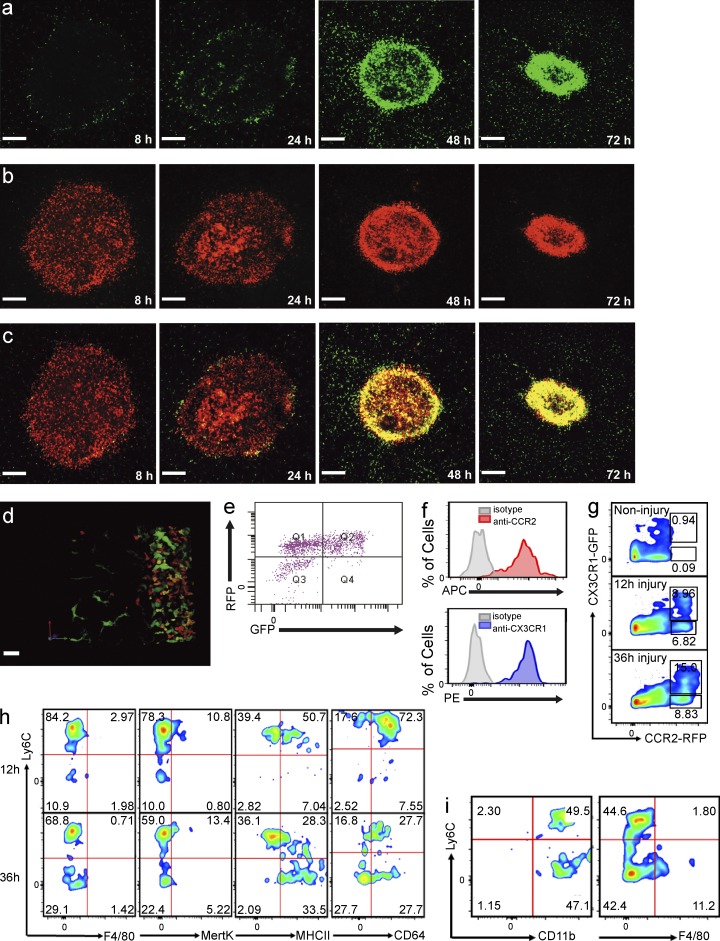
Figure 3.
CCR2+CX3CR1+ double-positive monocytes surround and enter the site of hepatic sterile injury. (a-c) Representative images taken 8 to 72 h after focal injury to the liver of Ccr2RFP/+/Cx3cr1GFP/+ mice. (a) GFP, (b) RFP, and (c) overlay. Bars, 200 µm. (d) Extended focus image generated from a z-stack of the monocytic ring surrounding a focal injury in the liver of a Ccr2RFP/+/Cx3cr1GFP/+ mouse demonstrating a spectrum of monocytes (red, orange, yellow, and green). (a–d) Images are representative of at least six independent experiments. (e) Representative FACS analysis of cells isolated from biopsy punches of the focal necrotic injury sites 48 h after injury, confirming the presence of a spectrum of monocytes. Pregated on size to exclude debris, viability, and CD11b+Ly6C+. (f) FACS analysis of cells obtained from the injury showing surface expression of CCR2 (top) and CX3CR1 (lower). Cells pregated on size, viability, Ly6G−CD45+, and RFP+GFP+ followed by measurement of APC-conjugated anti-CCR2 labeling (top) or pregated on size, viability, and Ly6G−CD45+, CCR2+GFP+, followed by measurement of PE-conjugated anti-CX3CR1 labeling (lower). (g) FACS analysis of CCR2+CX3CR1+ cells within the injury at the indicated time points. Cells pregated on size, viability, and Ly6G−CD45+. (h) FACS phenotyping of the CCR2+CX3CR1+ cells 12 and 36 h after focal liver injury. Cells pregated on size, viability, and Ly6G−CD45+. (i) FACS analysis of cells obtained 72 h after injury showing CD11b, Ly6C, and F4/80 expression. Cells pregated on size, viability, and Ly6G−CD45+RFP+GFP+. All FACS data are representative of at least three independent experiments.

