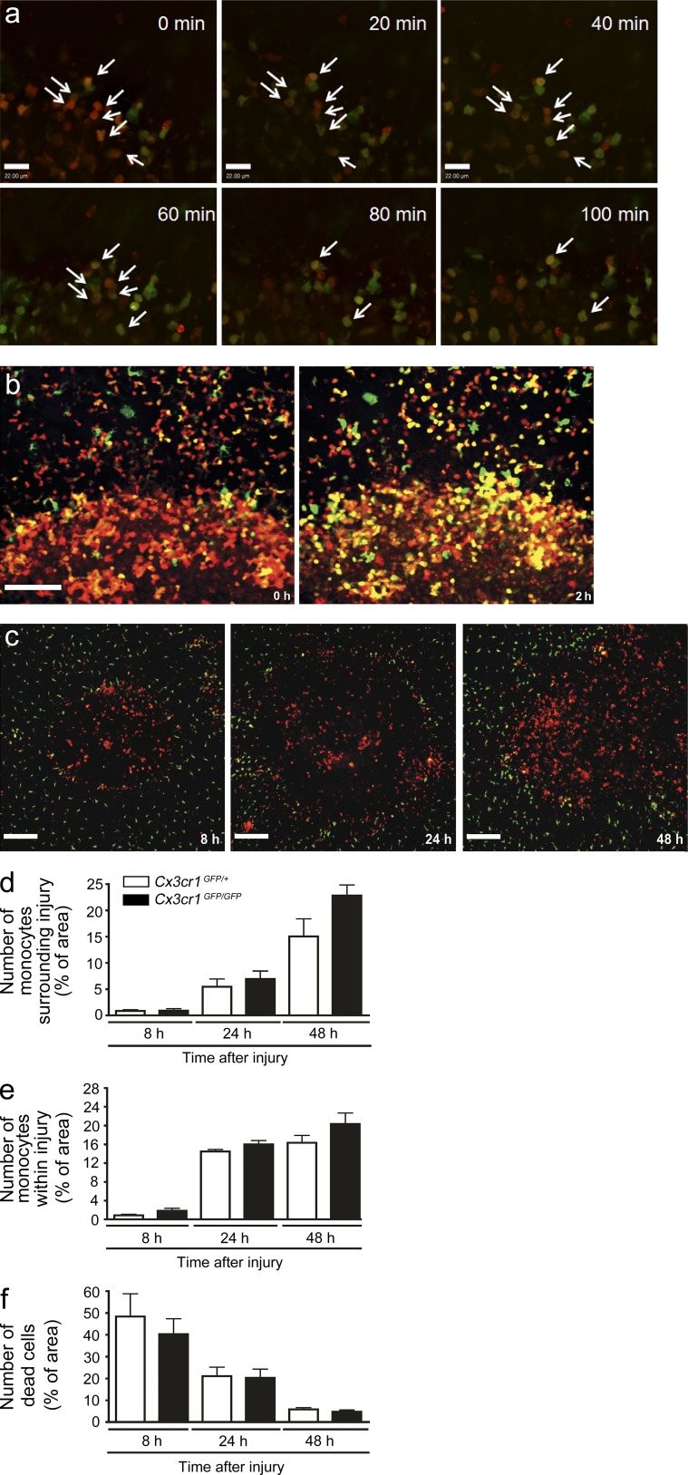
Figure 4.
Accumulation of reparative CX3CR1+ monocytes at the site of sterile injury is independent of the CX3CR1 receptor. (a) A 2 mm × 1 mm biopsy punch of the sterile injury site was harvested from a Ccr2RFP/+/Cx3cr1GFP/+ mouse 24 h after injury, maintained at 37°C, and 5% CO2 and imaged. Time-lapsed images demonstrate a shift from red to green in individual cells (arrows). Bar, 22 µm. Data are representative of two independent experiments. (b) Image of the injury border immediately after tissue harvest (left) and again after 2 h of culture at 37°C and 5% CO2 (right). Bar, 100 µm. Data representative of two independent experiments. (c) Representative images taken from 8 to 48 h after focal liver injury demonstrating failure to recruit Ccr2RFP/RFP cells to a site of injury results in the lack of appearance of GFP+ cells in Ccr2RFP/RFP/Cx3cr1GFP/+ mice. (d and e) Quantification of GFP monocytes in Cx3cr1GFP/+ and Cx3cr1GFP/GFP mice (measured by percentage of area covered by GFP) either surrounding lesion (d) or within the lesion (e). (f) Quantification of dead cells (measured by percentage of area covered by Sytox orange) within the lesion. (d–f) n = 6 mice per group; error bars are the SEM.

