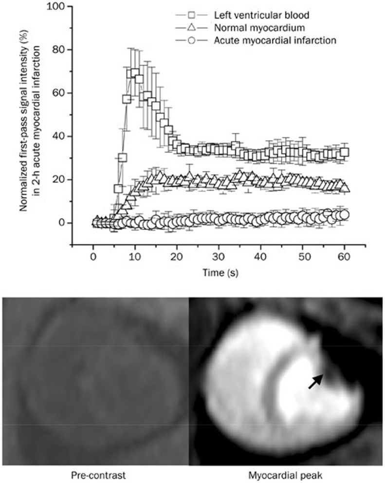
Figure 1.
Time intensity curves and representative images of first-pass perfusion MRI in acute myocardial infarction. The first-pass signal intensity in acutely infarcted myocardium did not change significantly, whereas the remote myocardium had a substantial signal elevation during the first pass of the contrast agent (upper panel). Therefore, first-pass perfusion MRI identified the acute myocardial infarction as a region of perfusion defect with hypoenhancement (arrowheads, lower panel).

