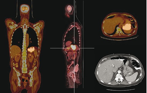
Figure 1.

Whole body positron emission tomography (18-FDG-PET/CT) scan identifying the location of the malignancy and demonstrating no pathological uptake in any other organs. Left: coronal section PET/CT scan identifying location of primary gastric cancer. Center: pathologic uptake of 18-FDG in primary gastric cancer, sagittal section. Top right: transverse PET/CT of primary gastric cancer. Bottom right: transverse section CT scan identifying large, ulcerated neoplasm.
