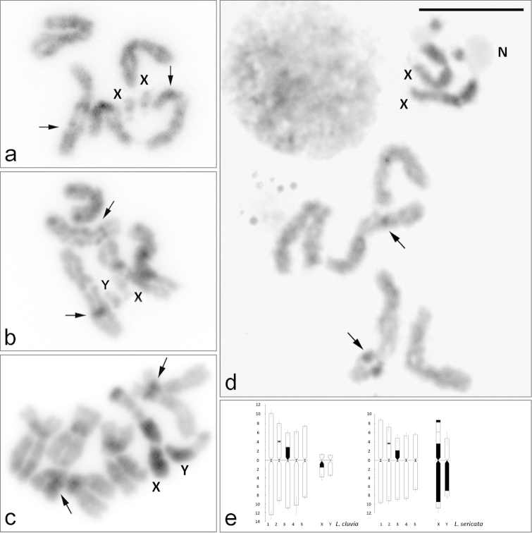
Figure 2.
C-banding on female and male mitotic chromosomes of Lucilia cluvia (a–b) and Lucilia sericata (c–d), stained with 3% Giemsa, and C-banded idiograms of autosomes and sex chromosomes of Lucilia cluvia and Lucilia sericata (e). X, Y = sex chromosomes. N = nucleolus. Arrows indicate C-positive heterochromatin bands at the secondary constriction in chromosome 2 and at interstitial position in chromosome 3. Bar = 10 μm.

