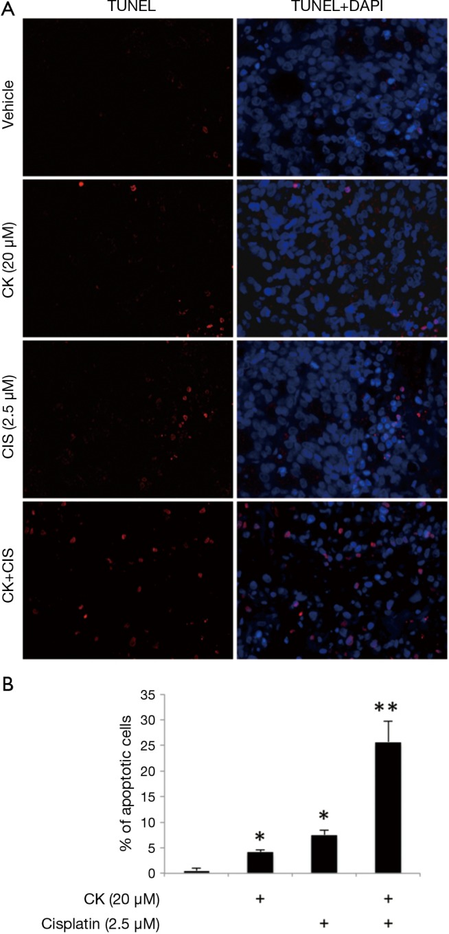
Figure 3.

Compound K synergizes cisplatin induced apoptosis. (A) Representative TUNEL staining pictures. H460 cells were treated with 20 µM CK and 2.5 µM cisplatin for 24 hr individually or in combination and TUNEL staining was conducted to detect cell apoptosis. DAPI was used to stain nuclei; (B) quantification of percentage of apoptotic cells shown in (A). Four fields from each treatment group were randomly chosen and TUNEL positive cells were counted and calculated as percentage of total cells. Data are presented as mean ± SD. *, P<0.05 from untreated control; **, P<0.01 from single treatment.
