Figure 2.

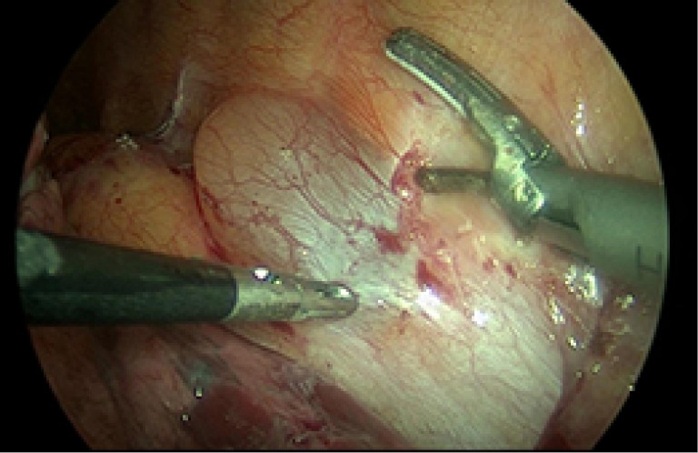
The thoracoscopic operative view showing the mass with a 3 mm camera (Storz) over the patient’s 6th thoracic vertebra abutting her aorta. The 5 mm grasper and 5 mm harmonic scalpel instruments are seen manipulating the cystic mass.

The thoracoscopic operative view showing the mass with a 3 mm camera (Storz) over the patient’s 6th thoracic vertebra abutting her aorta. The 5 mm grasper and 5 mm harmonic scalpel instruments are seen manipulating the cystic mass.