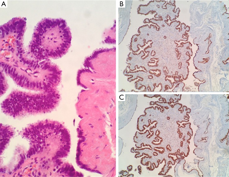
Figure 3.
(A) Ciliated columnar cells and flattened epithelium and surrounding smooth muscle bundles are seen in the cystic wall (Hematoxylin and eosin, 400×); (B,C) immunostaining shows positive staining for estrogen and progesterone receptors, respectively (estrogen receptor alpha and progesterone receptor, 100×).

