
Figure 2.
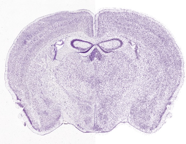
Predictive reconstruction of all cell positions in the whole mouse brain. The left side of the image shows a Nissl-stained section from the Allen Mouse Brain Atlas. The right side shows a digital reconstruction of the cell positions (neurons and glia) obtained algorithmically by analysing the data (C. Erö et al. 2014, unpublished data).