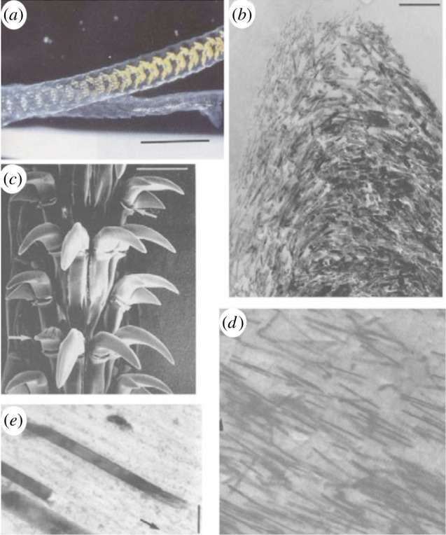
Figure 1.

Structure of the common limpet tooth (Patella vulgata). (a) Optical image of the tongue-like radula containing bands of teeth along a length of many centimetres. (b) Scanning electron micrograph of the teeth groupings with each tooth length approximately 100 μm. High-magnification electron microscopy images of the tooth cusp show (c) the changing orientation of the nanofibrous goethite in the chitin matrix and (d) the high anisotropy of the composite at the anterior and posterior edges owing to alignment of the goethite, note the mineral fibre length of approx. 3 μm, with (e) close-up of the tooth indicating the distinct phases of the goethite ‘reinforcing fibre’ and the chitin ‘matrix’ highlighting the structural resemblance to a fibre-reinforced composite material with an average fibre diameter of approx. 20 nm. Adapted from reference [12]. (Online version in colour.)
