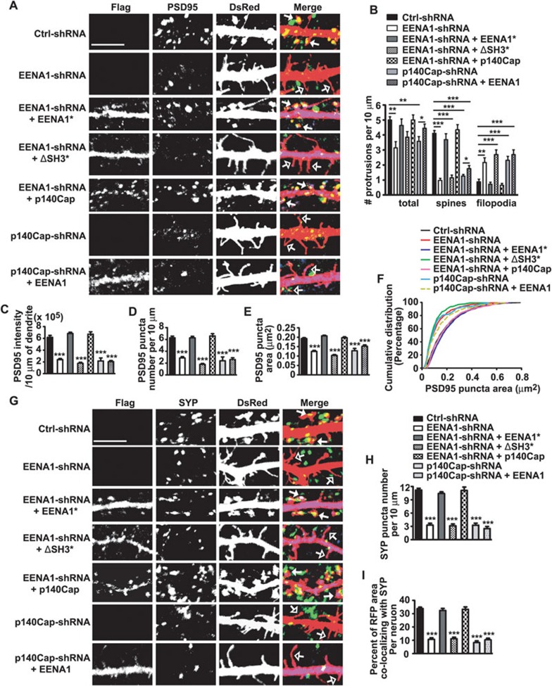
Figure 4.
Endophilin A1 regulates spine morphogenesis and synapse formation through its interaction with p140Cap. (A) Cultured hippocampal neurons were transfected with shRNA constructs or cotransfected with constructs encoding EENA1-shRNA and RNAi-resistant Flag-tagged endophilin A1, RNAi-resistant Flag-tagged endophilin A1 lacking SH3 domain (ΔSH3; indicated by asterisk), Flag-tagged p140Cap, or constructs encoding p140Cap-shRNA and Flag-tagged endophilin A1 at DIV16-17 followed by immunostaining with antibodies to PSD95 (green), Flag (blue) and DsRed at DIV21. Shown are representative confocal images. Filled arrows, spines; open arrows, filopodia. Scale bar, 5 μm. (B) Quantification of dendritic protrusion density of transfected neurons in A (number of cells analyzed, Ctrl-shRNA: 15, EENA1-shRNA: 15, EENA1-shRNA + EENA1*: 15, EENA1-shRNA + ΔSH3*: 18, EENA1-shRNA + p140Cap FL: 15, p140Cap-shRNA: 15, p140Cap-shRNA + EENA1: 18). More than 500 protrusions were analyzed for each group. All values are shown as mean ± SEM. Statistical test: ***P < 0.001, **P < 0.01, *P < 0.05; one-way ANOVA followed by Newman-Keuls multiple comparison post hoc tests. (C-E) Quantification of PSD95 intensity (C), puncta number (D) and puncta area (E) in dendrites of transfected neurons in A (number of cells analyzed, Ctrl-shRNA: 23, EENA1-shRNA: 25, EENA1-shRNA + EENA1*: 24, EENA1-shRNA + ΔSH3*: 22, EENA1-shRNA + p140Cap FL: 18, p140Cap-shRNA: 18, p140Cap-shRNA + EENA1: 28. number of puncta analyzed, Ctrl-shRNA: 990, EENA1-shRNA: 578, EENA1-shRNA + EENA1*: 1489, EENA1-shRNA + ΔSH3*: 449, EENA1-shRNA + p140Cap FL: 909, p140Cap-shRNA: 397, p140Cap-shRNA + EENA1: 432). More than 1 000 μm of dendrite length was analyzed for each group. All values are shown as mean ± SEM. Statistical test: ***P < 0.001; one-way ANOVA followed by Dunnett's multiple-comparison post hoc tests. (F) Cumulative distribution of PSD95 puncta area. (G) Cultured hippocampal neurons were transfected with shRNA constructs or cotransfected with constructs encoding EENA1-shRNA and RNAi-resistant Flag-tagged endophilin A1, RNAi-resistant Flag-tagged endophilin A1 lacking SH3 domain (ΔSH3; indicated by asterisk), Flag-tagged p140Cap, or constructs encoding p140Cap-shRNA and Flag-tagged endophilin A1 at DIV16-17 followed by immunostaining with antibodies to synaptophysin (SYP, green), Flag (blue) and DsRed at DIV21. Shown are representative confocal images. Filled arrows, spines; Open arrows, filopodia. Scale bar, 5 μm. (H, I) Quantitative analysis of the SYP puncta number along dendrites (H) or percentage of dendritic area colocalizing with SYP (I) (number of cells analyzed, Ctrl-shRNA: 16, EENA1-shRNA: 18, EENA1-shRNA + EENA1*: 15, EENA1-shRNA + ΔSH3*: 15, EENA1-shRNA + p140Cap FL: 16, p140Cap-shRNA: 13, p140Cap-shRNA + EENA1: 15). More than 600 μm of dendrite length was analyzed for each group. All values are shown as mean ± SEM. Statistical test: ***P < 0.001; one-way ANOVA followed by Dunnett's multiple-comparison post hoc tests.

