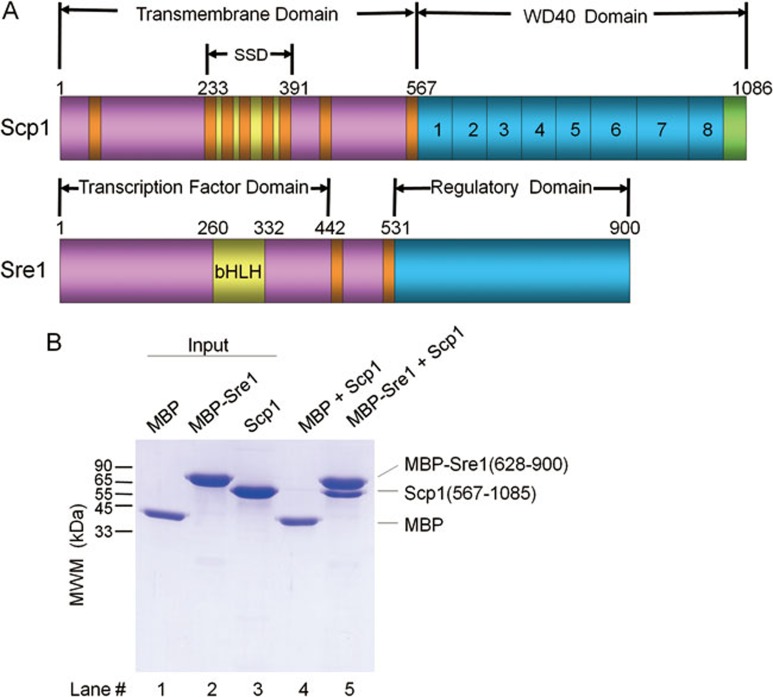
Figure 1.
In vitro reconstitution of the Sre1-Scp1 cytosolic complex. (A) Schematic illustration of the domain organizations of Scp1 and Sre1. Transmembrane helices are colored red and the carboxyl terminal tail of Scp1 is colored green. “SSD” stands for sterol sensing domain. The eight WD40 repeats of Scp1 are numbered 1-8. (B) Purified recombinant proteins of the C-terminal domains of Scp1 and Sre1 form complex in vitro. The MBP-fused regulatory domain of Sre1 (residues 628-900, and named MBP-Sre1) was immobilized on amylose resin to pull down Scp1-WD40 (residues 567-1 085). MBP was tested as negative control. Details of the experiments can be found in Materials and Methods.

