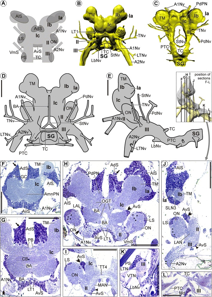
Figure 2.

Morphology of the brain in Mictocaris halope . Overview and semi-thin sections. A: Schematic depiction of our simplified nomenclature for soma clusters. B-E: Neuropil and nerves without somata. B, C: 3D-reconstructions in (B): anterior and (C): dorsal view. D, E: Schematic drawing in (D): posterior and (E): lateral view. F-L: Transverse semi-thin sections, ordered from anterior to posterior. F-H: Arrow points at a large dorsal extension of anterodorsal somata (AdS). G, H: Rocket points at the lateral root of the antenna 1 nerve (A1Nv). H: Points mark the olfactory glomeruli in the olfactory lobes (ON). H-J: Double arrowheads point at the large lateral extensions of lateral somata (LS). I-J: Simple arrowhead points at a large ventral extension of anteroventral somata (AvS). Scale bars: 50 μm.
