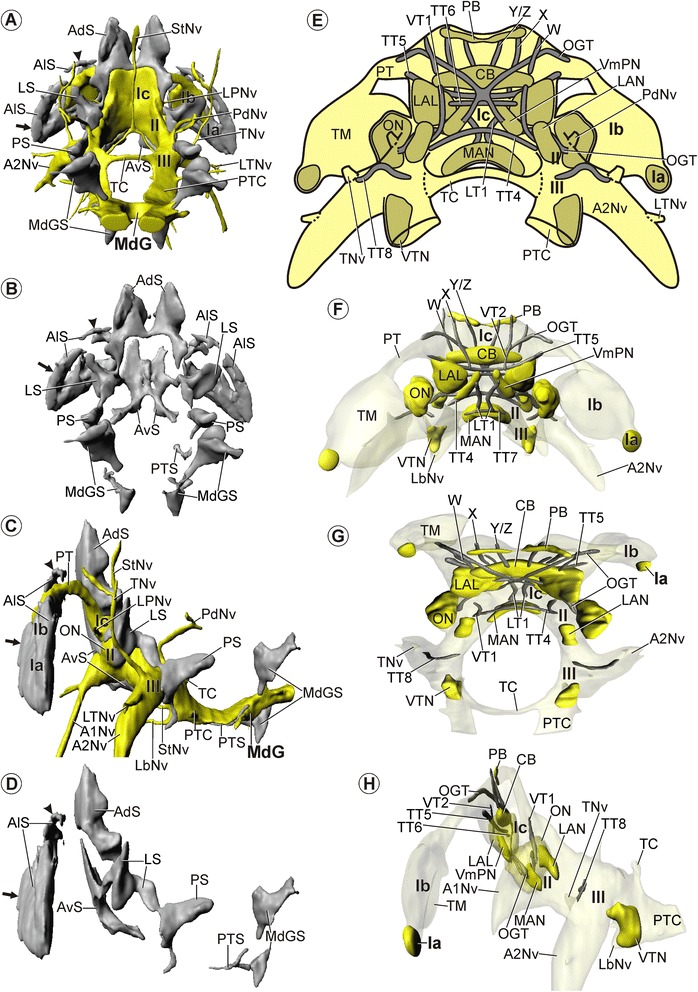
Figure 7.

Morphology of the brain in Spelaeogriphus lepidops . Soma clusters, neuropil, and internal structure – click on A and F for interactive 3D models. A, B: Soma clusters (grey) in posterior view (A): with neuropil (yellow) and (B): without neuropil. C, D: Soma clusters in lateral view (C): with and (D): without neuropil. A-D: Arrow points at a large cluster and arrowhead at a small cluster of anterolateral somata (AlS). E: Schematic drawing of neuropils (dark yellow) and tracts (grey) in posterior view. F-H: 3D-reconstructions of neuropils and tracts in (F): anterior, (G): dorsal, and (H): lateral view. Neuropils are in yellow; tracts are in grey. H: Lateral accessory lobe (LAL) and olfactory lobe (ON) is shown semitransparent. Brain width about 300 μm.
