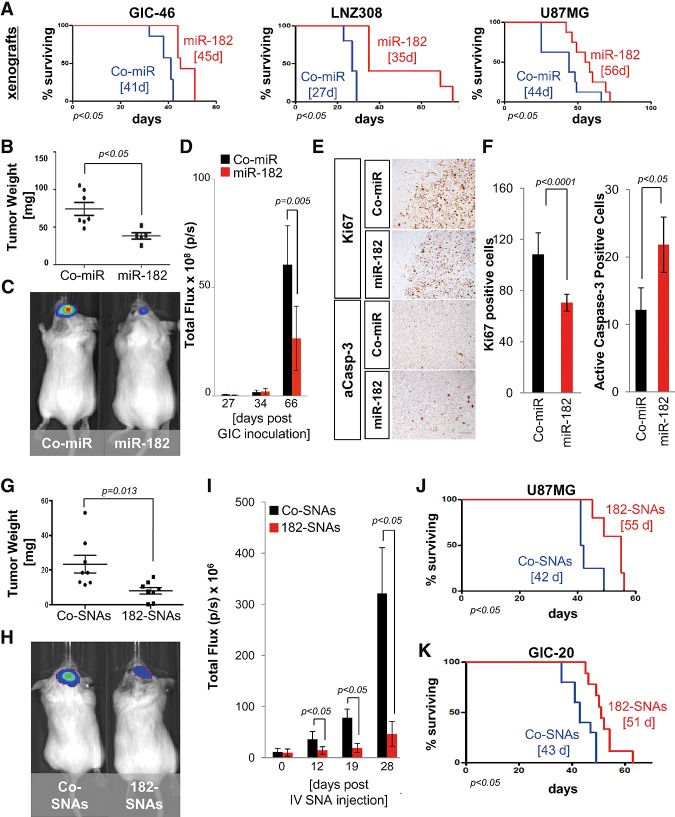
Figure 7.
182-SNAs reduce tumor growth in vivo. (A) Orthotopic xenograft survival analysis with glioma cells and GICs engineered to stably express miR-182 revealed that miR-182 expression increases survival of animal subjects. Median survival is indicated. (B–F) Analysis of tumor burden by weight (B) and bioluminescence imaging (C,D). (E) Ki67 and caspase-3 IHC in coronal brain sections of Co-miR-expressing and miR-182-expressing GIC-derived xenografts. Bar, 100 μm. (F) Quantification of Ki67 and caspase-3 in xenograft specimens. (G) Weight of U87MG-derived xenografted tumors extracted from SCID mice 21 d after intravenous treatment with Co-SNAs or 182-SNAs. (H) Bioluminescence imaging of xenograft tumors derived from GIC-20 12 d after intravenous treatment with Co-SNAs or 182-SNAs. (I) Quantification of bioluminescence signal up to 28 d after treatment with Co-SNAs or 182-SNAs. (J,K) Kaplan-Meyer survival curves of SCID mice carrying xenografted glioma tumors (U87MG and GIC-20), which were treated with intravenously administered Co-SNAs or 182-SNAs. Median survival is indicated.

