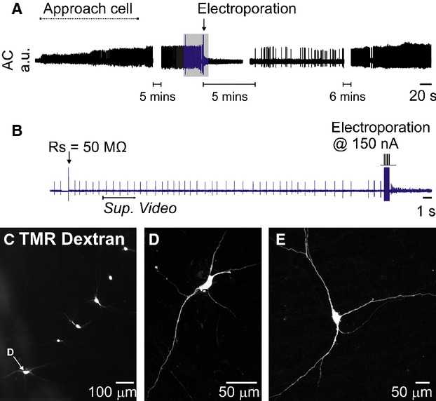
Figure 2.

Extracellular recording and constant‐current electroporation of a spontaneously active neuron in an acute brain slice (same recording as Video S1). (A) Overview of entire recording. (B) Detailed view of portion indicated in blue in A. To electroporate Rs was first measured using the amplifier bridge‐balance function and used to calculate the appropriate electroporation current (see Fig. 1). A 200 Hz train of 100 × 150 nA, 1 ms pulses immediately filled the cell with 1% tetramethylrhodoamine (TMR)‐dextran and abruptly halted its spontaneous discharge. Spontaneous activity returned after five minutes and was maintained for the remainder of the experiment. Sup. Video indicates starting point of Supplementary Video 1. (C) Low‐power fluorescence image of six dextran‐filled neurons recorded and electroporated in a single slice (D) Confocal image of the neuron indicated in C. (E) Example of a neuron from a different experiment.
