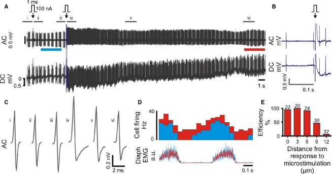
Figure 4.

Single‐cell microstimulation of a medullary respiratory neuron in vivo. (A) 100 nA microstimulation (arrow) initially evoked no effect on neuronal firing. The pipette was advanced 3 μm and stimulation repeated. This time the stimulus evoked transient stereotypical changes in firing frequency, spike amplitude and spike shape, apparent in the AC trace, and a small hyperpolarization of the pipette, visible as a 1 mV shift in the DC trace. (B) Expanded view of region drawn in blue in A. (C) Average spike waveforms; source data indicated in A. (D) Diaphragm‐triggered histograms of neuronal firing before (cyan, bar indicated in A) and after (red, bar indicated in A) microstimulation: the firing pattern is maintained over the recording. (E) Response to single‐cell microstimulation (0 μm) is correlated with high labeling efficiency (TMR‐dextran, in vitro), which decreases as the pipette is withdrawn from the cell membrane. Number of replicates shown over each series.
