
Figure 3.1.
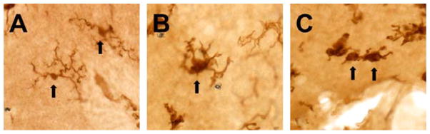
Microglial morphology exhibits a stepwise modification as the process of activation changes the functional phenotype of the cell. Microglial cells in the normal, unperturbed CNS exhibit a ramified morphology with a small soma, limited cytoplasm, and long, thin, highly branched processes (A). With activation, the cells hypertrophy and the processes retract, becoming broader and stunted with less branching (B). Highly activated cells progress toward an ameboid, hypertrophied form with few or no processes (C). These images are from the postnatal mouse cerebellum and are representative of the described morphological phenotypes of microglia. The microglial cells in these images were visualized with immunohistochemistry against the ionized calcium-binding adapter molecule (Iba-1), classically used to identify CNS microglia. Arrows indicate microglial cells with the described morphologies.