
Figure 1.
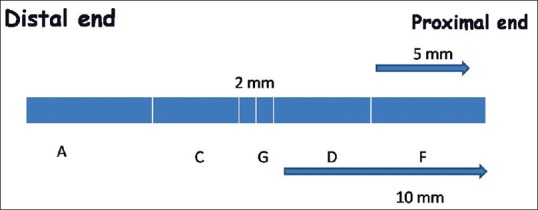
The schematic diagram of divisions of the harvested nerve for morphometric analysis. ‘G’ is the anastomosis site. ‘A’ is the distal most site while ‘F’ is the proximal site. Intermediate sites ‘C’ and ‘D’ respectively were also defined. Sections were taken from these sites in each specimen for analysis