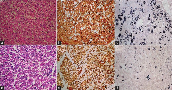
Figure 2.

(a) At the distal most site at 15 days, in the test group showing Schwann cell proliferation, degenerating axons, HE ×400. (b) Axons are small, haphazardly placed and sparsely distributed, NF ×400. (c) Extensive breakdown of myelin rings; all are thinly myelinated, Loyez ×400. (d) Extensive Schwann cell proliferation, most of the area showing degenerative changes, HE ×400. (e) In control cases very small axons seen with haphazard arrangement in most of areas, NF ×400. (f) extensive breakdown of myelin rings and majority are thinly myelinated, Loyez ×400
