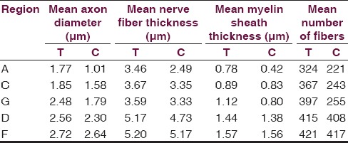
Table 4.
Shows morphometric analysis at 60 days along with statistical analysis. Axon diameter, myelin thickness and nerve thickness at distal sites were significantly more in the test group than the control group at 60 days (P<0.05)

Shows morphometric analysis at 60 days along with statistical analysis. Axon diameter, myelin thickness and nerve thickness at distal sites were significantly more in the test group than the control group at 60 days (P<0.05)
