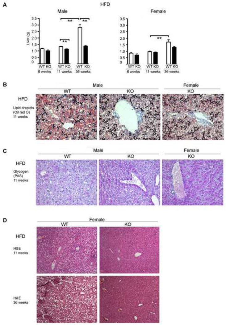
Figure 5. Cyp1b1-ko mice exhibit decreased liver steatosis, but increased glycogen retention.
A. Liver mass at 6, 11 and 36 weeks of HFD exposure in C57BL/6j (WT) and Cyp1b1-ko (KO) mice. The bar graphs indicate the average liver mass ± SEM for male WT (n=19, 6-week; n=22, 11-week; n=11, 36-week) and KO (n=4, 6-week; n=19, 11 week; n=6, 36-week) and female WT (n=13, 6-week; n=12, 11-week; n=4, 36-week) and KO (n=4, 6-week; n=10, 11-week; n=3, 36-week) mice.
B. Oil Red O staining for hepatic sinusoidal lipid droplets in representative WT and KO mice fed the HFD for 11 weeks.
C. Pas staining for hepatic sinusoidal glycogen granules in representative male WT and KO mice fed the HFD for 11 weeks (for expanded region see Figure S4A).
D. H&E stained liver sections of representative female WT and KO mice after 11 and 36 weeks of HFD exposure. Increased lipid droplets in WT livers are shown by lipid vacuoles.
*p<0.05, ** p<0.01.

