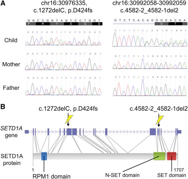
Figure 1. Two De Novo LOF Indels in SETD1A.
(A) Two de novo LOF indels in the SETD1A gene confirmed by Sanger sequencing. (B) Structure of the SETD1A gene and the SETD1A protein along with the positions of the two LOF indels. The de novo frameshift indel variant (D424fs) (left) creates an early stop codon and leads to protein truncation. The de novo indel variant c.4582-2_4582-1 del2 variant (right) changes the canonical splice acceptor site sequence adjacent to exon 16 from AG to GG.

