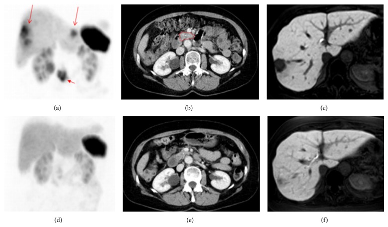
Figure 4.
Patient 5 before therapy ((a)–(c)) and after three cycles of 213Bi-DOTATOC ((d)–(f)) to a dose of 4 GBq. (a) Beta-resistant residuals in the liver (long arrows) and primary tumor (short arrow) are present in the 68Ga-DOTATOC PET maximum intensity projection image. (b) Contrast enhanced CT image with the primary tumor outlined in red. (c) In the MR image with hepatocyte-specific contrast medium, the liver metastases appear as black cavities against the enhancing normal liver parenchyma. ((d)–(f)) After three cycles of 213Bi-DOTATOC to a dose of 4 GBq, the lesions have diminished on the PET image (d) and CT image (e). Also on the MR image (f), the residual lesion has almost disappeared as shown by the growth of normal hepatocytes demonstrated by the uptake of the hepatocyte-specific contrast medium [36].

