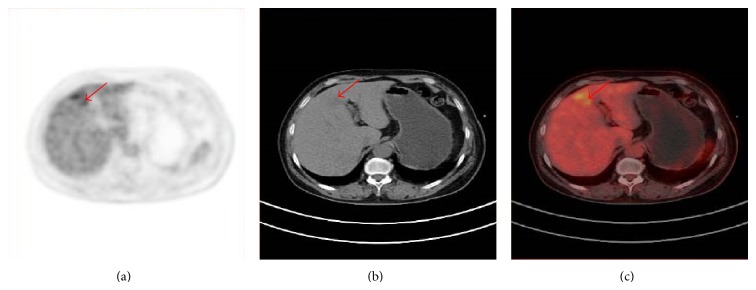
Figure 2.

PET, CT and PET/CT of a 59 year old patient, referred for restaging because of elevated CA 15-3 level 12 years after surgically resected breast carcinoma of the left breast. The PET demonstrates a focal increased FDG uptake in the liver (red arrow), which is considered as malignant. The CT shows a slightly low dense area in the left lobe of liver. The PET/CT is considered as suspicious because of the PET findings. Metastatic involvement of the liver was proved by follow-up.
