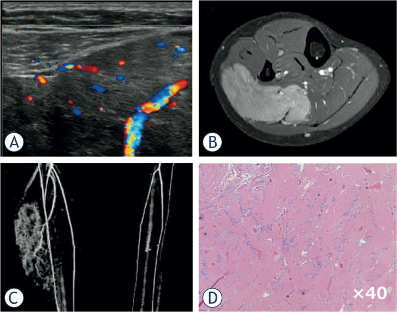
FIGURE 3.
Intramuscular haemangioma of gastrocnemius. A 19-year-old woman presented with a swelling and mild pain in her right lower leg. She experienced increased swelling and pain after exercise or long walks. (A) Ultrasound revealed an ill-defined mass with a mixed inner texture in the calf muscle. Colour Doppler examination showed multiple vessels within the tumour, corresponding to type IV in the classification described by Giovagnorio et al. (B) MRI showed a mass with an irregular border in the soleus muscle. (C) CT angiography revealed multiple vessels branching from the tibial artery. (D) Pathological analysis of a biopsy specimen demonstrated multiple vessels between the skeletal muscles with no atypia.

