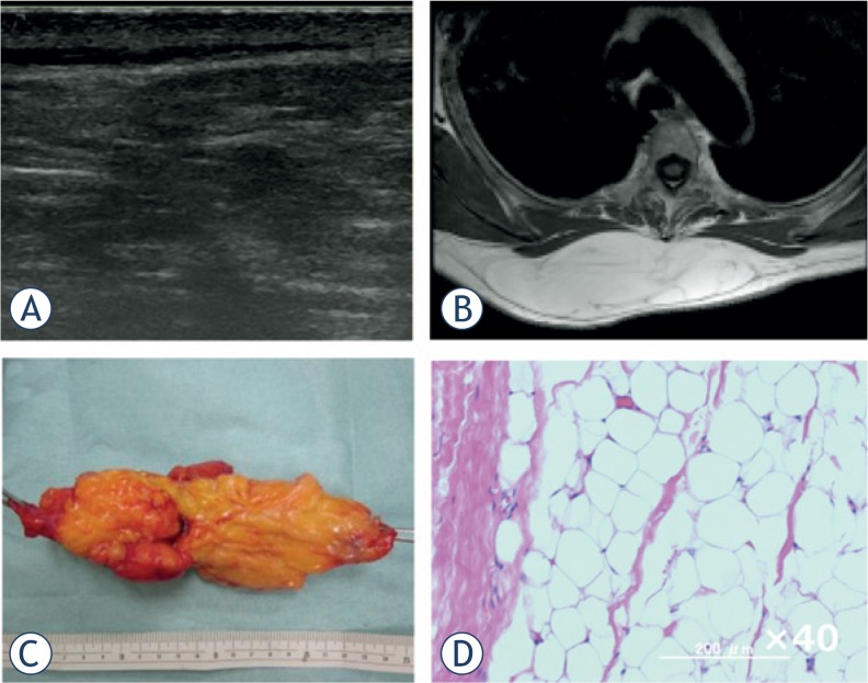
FIGURE 4.
Subcutaneous lipoma of the back. A 55-year-old woman presented with a subcutaneous mass on her back. (A) Ultrasound revealed a highly echoic mass with a mixed inner texture and no intratumoural vessels by Doppler echo. (B) MRI showed a high-intensity mass within the subcutaneous fat tissue on T1-weighted images. (C) Gross appearance of the marginally resected tumour. (D) Pathological examination of the tumour revealed mature adipocytes with no atypical cells.

