Figure 1.
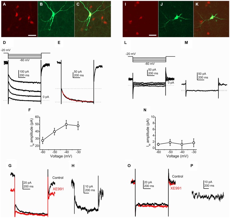
Cholinergic neurons possess M-current, whereas GABAergic neurons lack it. (A–C) Identification of a cholinergic neuron. Scale bar = 50 μm. (A) ChAT-dependent tdTomato expression (red). (B) Biocytin labeling of a recorded neuron (green). (C) Merged image. (D) Current traces from a cholinergic neuron elicited by the voltage protocol at the top of the panel. (E) Current trace at −40 mV repolarizing step. Dotted lines indicate the instantaneous (upper dotted line) and the steady state (lower dotted line) current components. The M-current was determined as the difference of these current components. The red trace indicates the fitting of the declining phase of the current (see text). (F) Voltage-dependence of the M-current amplitude (X axis: amplitudes of repolarizing current steps). (G) Pharmacological identification of the M-current (black = control; red = 20 μM XE991). (H) The XE991-sensitive current; calculated by the digital subtraction of the control and XE991-resistant current traces. The clear difference of XE991-sensitive current amplitudes recorded on −20 and −40 mV also represent the presence of M-current. (I–K) Identification of a GABAergic neuron. (I) GAD2-dependent tdTomato expression (red). (J) Biocytin labeling of a recorded neuron (green). (K) Merged image. (L) Current traces from a GABAergic neuron elicited by the same voltage protocol as on panel (D). (M) Current trace at −40 mV repolarizing step. Note that there is almost no difference between the instantaneous and steady state currents. (N) None of the repolarizing steps elicited M-current. (O) Currents from GABAergic neurons did not show XE991-sensitivity. (P) Digital subtraction of current traces under control conditions and in the presence of XE991. The lack of difference of XE991-sensitive current amplitudes recorded on −20 and −40 mV indicates that M-current was not recorded.

