
Figure 2.
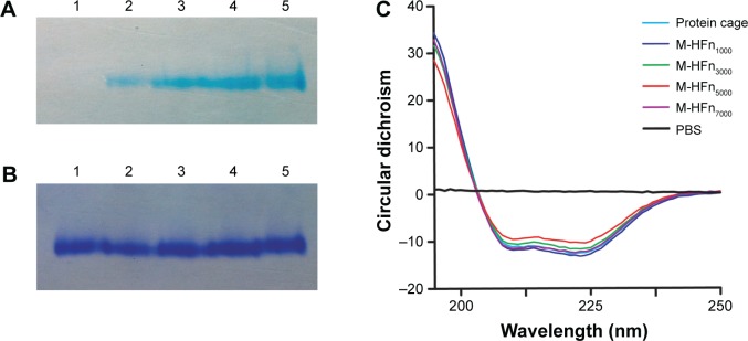
Native PAGE analysis of assembled HFn cage structure of M-HFn nanoparticles.
Notes: Gel was stained with (A) potassium ferrocyanide and (B) Coomassie Brilliant Blue R250. Lane 1, HFn cage; lane 2, M-HFn1000; lane 3, M-HFn3000; lane 4, M-HFn5000; and lane 5, M-HFn7000. (C) CD spectra determination of the secondary protein structural stability of M-HFn nanoparticles.
Abbreviations: HFn, H chain ferritin; M-HFn, ferrimagnetic H-ferritin; PAGE, polyacrylamide gel electrophoresis; CD, circular dichroism; PBS, phosphate-buffered saline.